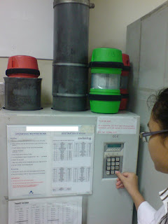
Before that, do you know how specimens reach the lab? All along I thought they were sent in by some medical staff every now and then. But one day when I was helping out at the reception, I realised it was not. The specimens are received in the lab via a pneumatic transport system (that looks like water pipes) that is linked from every single ward or clinic to the lab. Samples from the wards to be sent to the lab are placed into a capsule, which contains a micro-chip that is recognised by the system to direct its way to its destination. In every ward or clinic, there will be this cupboard that contains the ‘pipe’. Empty capsules with reports from the lab (if available) will then be sent back to the respective wards using the same system. But because there is only one lane for sending out, the destination code must be keyed to ensure that they are sent back to the correct place. Isn’t this interesting? The WHOLE hospital is connected by all these ‘pipes’.

Capsules arriving from wards
 Capsules waiting to be sent back to clinics/wards
Sending back capsules with reports through the one and only lane
(destination code keyed)
Ok, back to haematology. ESR (Erythrocyte Sedimentation Rate) is one of the most common tests performed in the haematology lab. There are many ways to test for the ESR. In my lab, the sedi-rate P4-Micro System is used. It measures the rate at which red cells fall in the first 50 mins when anti-coagulated blood is allowed to stand. Red cell sedimentation occurs in 3 stages: in the preliminary stage where aggregates form within a few minutes. This is followed by a period of time in which the sinking of the aggregates takes place at a constant speed. Finally, as the aggregated cells pack together at the bottom of the test tube, sedimentation rate slows down.
ESR is used to help diagnose conditions associated with acute and chronic inflammation. When inflammation is present in the body, certain proteins cause red blood cells to stick together and fall more quickly than normal to the bottom of the tube. These proteins may be produced when there an infection, autoimmune disease, or cancer. However, ESR is said to be nonspecific because increases do not indicate the exact site of inflammation or the causative agent. For this reason, a sedimentation rate is done in conjunction with other tests to confirm a diagnosis. Once a diagnosis has been made, a sedimentation rate can be done to help check on the disease or see how well treatment is working.
To perform ESR:
2. Then, a thin pipette is being pushed downwards into the tube, until the blood fills the whole pipette, indicated by the ‘0’ marking.
3. Next, the tube with pipette will be allowed to stand.
4. Exactly after 50 mins, the number of mm the red cells fallen would be read.
5.Results are then recorded in the ESR record book and the request form, and entered manually into the LIS.
 Source taken from: http://www.globescientific.com/sedirate™-autozero-westergren-esr-system-3469-pi-574.html
Foetal RBCs contain mainly HbF (a2g2). They resist acid elution more than that of adult RBCs, which contain mainly HbA (a2b2). With this principle, foetal cells can take up the eosin when counterstained and appear as darkly stained red cells. On the other hand, adult cells will be disintegrated by the acid and therefore will appear as ghost cells (because the cells are dissolved, there is no more cells present to take up the stain).
To perform the KB test:
10 comments:
August 8, 2008 at 11:16 PM
Hello kahang!
The KB test you described sounds interesting. We studied before right? Anyway, why do you have to take the 3 differnt samples for the 3 different smears? How can the male blood act as a control?
Thanks!
Leslie
August 9, 2008 at 2:16 PM
Hello!!
You mentioned that fetal RBCs will resist acid elution more than adult RBCs. Why is that so? Is it because of the HbF? If it is, then how does it affect the RBCs' ability for acid elution?
Thanks!!
-Li Ping-
TG o2
August 9, 2008 at 3:44 PM
Hi KaHang!
Hope you are doing well. And ya, the capsule system is so cool. Ours uses the traditional box that travels above your head. HAha
Btw,in KB, How different are the cord blood and male male as positive and negative controls under microscopic examination?
And for the acid elution, is it affected by the type of Hb? As in HbF resist, HbA does not. If that's the case, is there any exceptional cases (conditions) where the adult cells do not disintergrate?
Thanks!
Ying Chee
TG01
August 10, 2008 at 2:46 AM
hi kahang,
for acid elution, may i know what acid is used? and for the controls for KB test? why must blood be taken from a male? instead of a female? thanks.
Malerie
TG02
August 10, 2008 at 11:10 PM
Hey Ka Hang,
here's a little question XD
You mentioned that," In this case, the test checks whether the baby’s blood had entered the maternal circulation. If positive.."
May i know how exactly does KB check for the above scenario.
And how to does it determine whether if it's +ve or -ve. Thanks !
Albert Chan
0604524I
August 11, 2008 at 10:47 AM
hey.
wgen you mention "ghost cells", do you mean they just die? or that it kinda just loses it colour under the microscope?
and just to clarify, light microscope will do right? its fine to identify the stains using the light microscope?
thanks(:
glad
August 15, 2008 at 12:40 AM
Hey Ka Hang~
Liyana here.. the explanation about the KB tests for foetal anaemia and Rh(+)/(-) has been clear (and your answers + pictures have been helpful!), but there's a but!
(hehe..)
How does the KB test help detect down syndrome or any other genetic disorders? KB test is mainly staining right.. i dont think it stains genes or chromosomes..?
Nor Liyana
0607927A
August 16, 2008 at 1:31 AM
Hi Liyana,
KB test does not detect chromosomal diseases. To detect chromosomal dieseases, the foetus's blood needs to be analysed before it is born. So the foetus's blood needs to be taken through the mother's tummy. In this way there is a possibility that the blood taken might be that of the mother's (the doctor may aim wrongly). Therefore, to confirm that the blood taken is from the foetus, KB test is performed.
If the test result is positive, this means that the blood taken is indeed from the foetus and this blood sample will be sent to the cytogenetics lab for chromosomal analysis.
Thanks.
Ka Hang
TG02
August 17, 2008 at 9:21 PM
Hi Kahang,
How do you all determine how much foetus's blood has went into the mother's circulation?
Jean Leong
TG02
August 20, 2008 at 9:41 PM
Hi Jean,
If foetal cells are detected, the number of foetal cells seen in 2000 adult cells and the volume can be deduced using the 'Mollison Calculation Formula' (it's rather chim, so i didnt mention it in my post):
2000/(2000 adult cells/# of fetal cells)
= 2400 x (# of fetal cells/2000)
= 1.2 x # of fetal cells counted in 2000 RBCs
= volume of fetal blood in maternal circulation (ml)
Thanks.
Ka Hang
TG02
Post a Comment