Replies to comments on Kahang's (Week 7) Post

Hi, I shall answer all your questions here:

To Leslie, Ying Chee & Malerie
I mentioned that 3 different blood smears will be made, one using the patient’s (women’s), one using a male’s and another with cord blood. Remember this test is to test for the presence of fetal cells in the mother’s blood? So the most important factor would be the fetal cells.

Cord blood from newborns is used as a positive control as it definitely contains fetal cells. When stained, they will take up the eosin counterstain and appear as pinkish red intact cells. If red cells are not seen in the positive control, it might be an indication that the stain is not working well.

Male’s blood is used as a negative control because it is impossible that fetal cells will be present. Normal cells of adults will be dissolved after the acid elution staining and appear as ghost cells (illustrations are provided below). This is done to show that the procedures for this test are carried out correctly. If fetal cells are seen in the male’s smear, cross contamination might have occurred or there were errors in the procedures.

In fact the negative control may not necessarily be taken from a male. It can also be taken from a non-pregnant female, as long as it is from an adult. But in certain cases where women are unaware of their pregnancy, the results from the negative control may reflect as positive (presence of fetal cells). So to be more accurate, my lab uses a male’s sample as the negative control.


To Li Ping & Ying Chee
Yes it’s the HbF that affects the resistance to acid elution. The hemaglobin F does not dissolve in the presence of acid and thus takes up the eosin counterstain and appear as red. The adult hemoglobin A cells are already dissolved when acid is introduced, therefore they appear as ‘ghosts’. The reason behind this resistant is not really known. I tried consulting the medtechs in my lab, they are not sure either, just said that it’s definitely related to the HbF component.


To Malerie
The acid elution solution consists of solutions A, B and C:

Solution A – 0.75% Haematoxylin + 96% Ethanol
Solution B – 2.4g of Ferric Chloride + 0.5% Hydrochloric Acid
Solution C – 80% Ethanol

Solutions A, B and C are mixed in the ratio 2:1:1 and this working solution must be prepared fresh.


To Albert, Ying Chee & Gladys
Gladys, yes light microscope would be sufficient. Slides are examined under x40 objective. If fetal cells are observed, counting is performed under oil immersion (x100 objective).

Microscope examination of:

Negative control – Normal adult cell contents are dissolved, therefore nothing can take up the eosin stain, cells appear as ‘ghost’ (cells only have its border with no content)

Positive control (cord blood) – All cells take up the eosin stain and appear as pinkish red intact cells

Positive Result – Stained red cells denote the presence of fetal cells.


(All pictures taken from lab's SOP, with permission from medtech)


Ka Hang
TG02

Answers for 'Week 5 and Earlier Sharings' queries

Hi everyone,

I hope you guys are doing well, in SIP and as well as in revising (!!) our other subjects. Hope You are in the pink of health too.. and unfortunately, I am not. haha.. it's the flu season again, i think~

I am here to answer some of the questions posed by some of you:

I will start with Sharon's -

How frequently do you clean CO2 cylinder and waterbath?

I hope you mean 'CO2 incubator', because I definitely do not clean any CO2 cylinder. >.^

- oops! I have just realised I typed 'CO2 cylinder' in my previous post~ Okay, am correcting it now -

The CO2 is stored in cylinders, and these cylinders (and the CO2) are supplied by Singapore Oxygen Air Liquide Pte Ltd. I place order for new supply/ies of CO2 every week, because the gas runs out every 7-9 days.

How frequent the cylinder has to be changed/ordered depends on 1. the amount of growing cells/microorganisms kept in the CO2 incubator, and 2. how frequent the gas leaks out of the incubator.

Say, for example, if I keep opening and closing the incubator, or open it for too long, more CO2 will be lost. So, yes, I have to be mindful when I am using the CO2 incubator.

The CO2 incubator is cleaned every month. This is to ensure no bacteria or any other microorganisms, that can contaminate the cell culture, grow in it. It will be a HUGE problem if there are contaminants! Imagine, you keep 20 flasks of rare cells in the incubator, and suddenly, they all died! And... it turns out that some fungi managed to sneak into the flasks and eat up all the nutrients in the media. nightmare!

Here is a better description of how important keeping the CO2 incubator clean is:

Cleaning and Disinfecting CO2 Incubators


A CO2 incubator is a basic piece of equipment in any laboratory involved in cultivation of in vitro cells. The culture bottles must be kept slightly opened in order to allow the penetration of CO2, and enable the formation of a bicarbonate - CO2 buffer system, which is the most common buffer system for tissue culture. Leakage of medium from the culture bottles, high humidity, suitable temperature, the water tray and the air flow, all form a fertile breeding ground for the development of contaminants in the incubator, which are liable to contaminate the tissue culture in all the semi-opened culture bottles.

As soon as contamination appears in the incubator, it is very difficult to eliminate it, and severe damage can be caused. Therefore, it is essential to systematically clean and disinfect the incubator. It is recommended to disinfect every 14 days, or once a month at the very least.

Retrieved August 11, 2008, from Biological Industries website

Firstly, I take out the tray (one at a time), place it at the sink and run DI water over it. Then, I spray 70% alcohol on the tray and wipe/dab with tissue paper. All these must be done quickly, to minimise the tray's exposure time to the surrounding air (i.e. dust may settle on the tray).

After all the trays and incubator wall (just spray with 70% alcohol) have been cleaned, the last item is the water tray, which should be cleaned and then filled with sterile water. What is the water used for? It is to maintain humidity inside the incubator.

To be safe, my lab uses Aquaguard 1 solution, a disinfectant from Biological Industries, to disinfect the water in the tray. 50mL of Aquaguard 1 to every 5L of sterile water.

Okay, as for the waterbath, the water has to be changed every 4 or 6 weeks. DI water, to be exact. We use Aquaguard 2 solution to disinfect the water. 2mL of Aquaguard 2 to every litre of water.

What is the difference between Aquaguard 1 and 2?

Simple: Aquaguard 1 is especially for CO2 incubator, while Aquaguard 2 is for waterbath. This is what stated at the supplier's website. It does not state the composition though.

Okay, hopefully this answers your question, Sharon. Do drop me a comment (or sms, even better! hehe) if you have any other doubts. See you this Friday, ya? ^_^


I-am-at-TP-library-going-home-soon,
Nor Liyana
0607927A

Week 7

Hi all, for the past 3 weeks, I was attached to the haematology lab. Like other clinical labs, it operates 24 hours and is very busy almost everyday. The most common test ordered is full blood count, but since sofie has already touched on it, I shall talk about other tests.

Before that, do you know how specimens reach the lab? All along I thought they were sent in by some medical staff every now and then. But one day when I was helping out at the reception, I realised it was not. The specimens are received in the lab via a pneumatic transport system (that looks like water pipes) that is linked from every single ward or clinic to the lab. Samples from the wards to be sent to the lab are placed into a capsule, which contains a micro-chip that is recognised by the system to direct its way to its destination. In every ward or clinic, there will be this cupboard that contains the ‘pipe’. Empty capsules with reports from the lab (if available) will then be sent back to the respective wards using the same system. But because there is only one lane for sending out, the destination code must be keyed to ensure that they are sent back to the correct place. Isn’t this interesting? The WHOLE hospital is connected by all these ‘pipes’.

Capsules arriving from wards


Capsules waiting to be sent back to clinics/wards
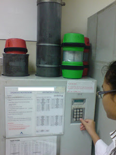

Sending back capsules with reports through the one and only lane
(destination code keyed)


Ok, back to haematology. ESR (Erythrocyte Sedimentation Rate) is one of the most common tests performed in the haematology lab. There are many ways to test for the ESR. In my lab, the sedi-rate P4-Micro System is used. It measures the rate at which red cells fall in the first 50 mins when anti-coagulated blood is allowed to stand. Red cell sedimentation occurs in 3 stages: in the preliminary stage where aggregates form within a few minutes. This is followed by a period of time in which the sinking of the aggregates takes place at a constant speed. Finally, as the aggregated cells pack together at the bottom of the test tube, sedimentation rate slows down.

ESR is used to help diagnose conditions associated with acute and chronic inflammation. When inflammation is present in the body, certain proteins cause red blood cells to stick together and fall more quickly than normal to the bottom of the tube. These proteins may be produced when there an infection, autoimmune disease, or cancer. However, ESR is said to be nonspecific because increases do not indicate the exact site of inflammation or the causative agent. For this reason, a sedimentation rate is done in conjunction with other tests to confirm a diagnosis. Once a diagnosis has been made, a sedimentation rate can be done to help check on the disease or see how well treatment is working.

To perform ESR:

1. 320µl of blood is transferred into an aquisel tube, which contains 0.08ml of sodium citrate.
2. Then, a thin pipette is being pushed downwards into the tube, until the blood fills the whole pipette, indicated by the ‘0’ marking.
3. Next, the tube with pipette will be allowed to stand.
4. Exactly after 50 mins, the number of mm the red cells fallen would be read.
5.Results are then recorded in the ESR record book and the request form, and entered manually into the LIS.


Another special test that I think relates to what we have learnt is the Kleihauer Betke Test (KB), which is used to detect the presence of foetal RBCs in the mother’s blood using the principle of acid elution (dissolving of cells). KB Test can be used to assess 3 conditions.

One of which is when a newborn baby is found to be anaemic. In this case, the test checks whether the baby’s blood had entered the maternal circulation. If positive, the amount of blood must be determined and from there, the doctor will be able to decide how much blood should be transfused.

The second condition is, when the mother is Rh(-ve) and baby is Rh(+). If the KB test reflects a positive result, rhogam must be administered within 72 hours to neutralize the foetal RBCs.

Also, KB test plays an important part in cytogenetic analysis. To detect genetic abnormalities such as down syndrome, blood from unborn foetus must be analysed. To facilitate the process, the foetus’s blood must be taken through the mother’s tummy. The KB test will confirm whether the taken blood belongs to that of the foetus and not the mother.

Foetal RBCs contain mainly HbF (a2g2). They resist acid elution more than that of adult RBCs, which contain mainly HbA (a2b2). With this principle, foetal cells can take up the eosin when counterstained and appear as darkly stained red cells. On the other hand, adult cells will be disintegrated by the acid and therefore will appear as ghost cells (because the cells are dissolved, there is no more cells present to take up the stain).

To perform the KB test:

1. 3 different smears (patient’s blood, blood of a male and cord blood) are first made. Cord blood smear act as the positive control, blood smear from male acts as the negative.
2. Slides are air dried.
3. Slides are fixed in 80% ethanol for 10 mins then stained using the acid elution method.
4. This is followed by counterstaining with eosin for 3 mins and then air dried.
5. Finally slides are examined under the microscope at high power.

The proportion of foetal RBCs that appear as pinkish red intact cells to that of adult RBCs (appear as ghost cells) is assessed in several fields. If foetal cells are detected, report the number of foetal cells seen in 2000 adult cells and the volume will be calculated.


Ka Hang
TG02